Publications
Evaluating the Translation Value of Two In Vivo Models for Breast Cancer Brain Metastases
Cancers (2026)
Abstract

In Vivo Studies of [161Tb]Tb-Trastuzumab Radiopharmaceutical Therapy in Human Epidermal Growth Factor Receptor 2–Expressing Breast Tumors Show High Tumor Uptake and Tumor Growth Suppression
, , , , , , , and
Abstract
Human epidermal growth factor receptor 2 (HER2) has emerged as attractive for targeted radionuclide therapy of HER2-expressing breast cancer. The present study aimed to investigate the image-derived biodistribution, tolerability, and efficacy of [161Tb]Tb-trastuzumab as a potential therapeutic alternative or supplement to trastuzumab and trastuzumab deruxtecan in HER2-expressing tumors. Methods: Trastuzumab was functionalized with DOTA by non–site-specific conjugation and radiolabeled with 161Tb. In vitro assays were performed to evaluate binding affinity and immunoreactivity of [161Tb]Tb-trastuzumab. The influence of mass dose on the biodistribution of [161Tb]Tb-trastuzumab was assessed in mice bearing subcutaneous BT474 tumors by SPECT/CT imaging performed 4, 24, 72, and 168 h after injection of 20 MBq of [161Tb]Tb-trastuzumab labeled at 3 specific activities. Tolerability was evaluated with escalating doses of 5, 10, and 20 MBq of [161Tb]Tb-trastuzumab, and the efficacy of [161Tb]Tb-trastuzumab was compared with that of the vehicle, trastuzumab, and trastuzumab deruxtecan groups. Results: Functionalization and radiolabeling resulted in a radiochemical yield of 97% ± 2% and a radiochemical purity above 99%. The binding affinity of functionalized and native trastuzumab was 0.83 and 0.39 nM, respectively, with an immunoreactive fraction of 89%. SPECT/CT imaging demonstrated peak tumor uptake 72 h after injection, with tumor uptake of 10.8 ± 1.1, 12.8 ± 2.2, and 6.3 ± 0.03 %ID/g of tissue for mice dosed with 0.5, 0.05, and 0.005 MBq/µg, respectively. Radiotoxicity was evident in mice dosed with 20 MBq of [161Tb]Tb-trastuzumab, whereas 5 and 10 MBq of [161Tb]Tb-trastuzumab were well tolerated. Antitumor efficacy was observed in mice dosed with 10 MBq of [161Tb]Tb-trastuzumab, which presented tumor volumes statistically significantly smaller than those of the vehicle group 104 d after treatment (P = 0.0007). Conclusion: Functionalization and radiolabeling did not affect binding affinity. Biodistribution of [161Tb]Tb-trastuzumab revealed high tumor uptake of more than 10 %ID/g of tissue, peaking at 72 h. [161Tb]Tb-trastuzumab in doses of up to 10 MBq was tolerated and highly effective at inhibiting tumor growth (P = 0.0007). These results support the potential of [161Tb]Tb-trastuzumab as an attractive treatment option for HER2-expressing cancers.

A novel synergistic drug combination of a mitogen-activated extracellular signal-regulated kinase inhibitor with [177Lu]Lu-rhPSMA-10.1 for prostate cancer treatment: Results of a preclinical evaluation
Nuclear Medicine and Biology (2025)
, Bart Cornelissen, , Freja Pretzmann, , Mathias Wikke Hallund, and
Abstract
The prostate-specific membrane antigen (PSMA)-targeted radiohybrid ligand [177Lu]Lu-rhPSMA-10.1 is a promising next-generation radiopharmaceutical therapy in prostate cancer. This preclinical evaluation comprised an in vitro screen of potential novel synergistic drug combinations with [177Lu]Lu-rhPSMA-10.1, and an in vivo efficacy analysis of the lead drug combination in PSMA-expressing prostate cancer xenografts. Methods: In total, 177 anticancer drugs were screened in a clonogenic survival assay of 22Rv1 cells which used 5-fold serial dilutions of the test drug (≤ 20 μM) to determine the half-maximal inhibitory concentration (IC50), compared to incubations of the test drug plus [177Lu]Lu-rhPSMA-10.1 (15 MBq) after 10 days. A subsequent focused screen assessed the impact of [177Lu]Lu-rhPSMA-10.1 (0–25 MBq/mL) on drug IC50. Synergy scores were determined using the zero interaction potency (ZIP) reference model (ZIP scores >5 % indicate high synergistic potency) and the multidimensional synergy of combinations (MuSyC) platform (log α >0 indicates synergistic potency). Therapeutic efficacy of the lead drug combination was evaluated in vivo: intravenous [177Lu]Lu-rhPSMA-10.1 (30 MBq, single dose) and oral cobimetinib (0.25 mg/day for 21 days) (alone/in combination) were administered to 22Rv1 tumor-bearing NMRI nude mice (eight mice/group plus untreated controls). Tumor volume was measured twice weekly for 69 days (two-way ANOVA and Tukey’s multiple comparisons test: data analyzed until three mice/group remained). KaplanMeier Log-rank survival analyses were performed. Results: In vitro screening identified cobimetinib (a mitogen-activated extracellular signal-regulated kinase inhibitor) as a lead candidate for synergistic combination with [177Lu]Lu-rhPSMA-10.1 across a wide concentration range (ZIP score=13 %). MuSyC analysis suggested synergistic efficacy from enhanced potency of both drugs in the combination (both log α>3). Combination treatment significantly suppressed tumor growth in vivo versus untreated controls (from Day 13–30; p<0.01) and [177Lu]Lu-rhPSMA-10.1 (from Day 17–30; p<0.001). Median survival was significantly longer with combination treatment (49 days) versus untreated controls (23 days; p=0.001) and [177Lu]Lu-rhPSMA-10.1 monotherapy (36 days; p=0.002). No major compound-related toxicity for cobimetinib ± [177Lu]Lu-rhPSMA-10.1 was observed. Conclusions: The combination of cobimetinib and [177Lu]Lu-rhPSMA-10.1 demonstrated enhanced preclinical therapeutic efficacy versus single agents, supporting clinical investigation of this novel drug combination in prostate cancer.

Preclinical Evaluation of 177Lu-rhPSMA-10.1, a Radiopharmaceutical for Prostate Cancer: Biodistribution and Therapeutic Efficacy
, , , , , and
Abstract
177Lu-rhPSMA-10.1 is a novel radiohybrid prostate-specific membrane antigen (PSMA)–targeted radiopharmaceutical therapy for prostate cancer. We conducted preclinical analyses on non–tumor-bearing BALB/c mice and on prostate cancer human xenograft mouse models (LNCAP and 22Rv1 xenografts) to evaluate its biodistribution and therapeutic efficacy. Methods: Longitudinal biodistribution of 177Lu-rhPSMA-10.1 was evaluated in BALB/c mice and 22Rv1 xenografts. Tissues of interest were harvested, and radioactivity was measured 1–168 h after injection of 1 MBq of 177Lu-rhPSMA-10.1 (4 per time point). Longitudinal biodistribution was compared with 177Lu-PSMA-I&T (1 MBq) in BALB/c mice and at a single time point (15 h) in 22Rv1 xenografts. The therapeutic efficacy of a single administration of 15, 30, or 45 MBq of 177Lu-rhPSMA-10.1 in LNCaP xenografts and 30 MBq of 177Lu-rhPSMA-10.1, 177Lu-PSMA-617, or 177Lu-PSMA-I&T in 22Rv1 xenografts (8 per group) was evaluated. Efficacy versus vehicle was evaluated on the basis of relative tumor volume and survival up to 49 d after administration. Statistical significance was evaluated with t testing (biodistribution data), 2-way repeated-measures ANOVA (tumor volume [analyzed until 3 per group remained]), or Kaplan–Meier log-rank analyses (survival). Results: Biodistribution of 177Lu-rhPSMA-10.1 in the BALB/c and 22Rv1 xenografts showed rapid clearance from blood and other normal tissues within 48 h, with the kidney showing the highest normal-organ uptake. Kidney uptake and retention were lower for 177Lu-rhPSMA-10.1 than for 177Lu-PSMA-I&T (6.5-fold lower at 12 h in BALB/c mice and 6.4-fold lower at 15 h in 22Rv1 xenografts; P < 0.01). High and sustained 177Lu-rhPSMA-10.1 tumor uptake was observed in 22Rv1 xenografts. This uptake was 2.3-fold higher than that of 177Lu-PSMA-I&T (15 h; P < 0.05). When efficacy was evaluated, 177Lu-rhPSMA-10.1 significantly suppressed tumor growth versus vehicle from day 11 (P < 0.05) in LNCaP xenografts in a dose-dependent manner and from day 18 (P < 0.05) in 22Rv1 xenografts and significantly prolonged median survival versus vehicle in both models. In 22Rv1 xenografts, 177Lu-rhPSMA-10.1 suppressed tumor growth versus vehicle to a greater extent than did 177Lu-PSMA-I&T (significant growth inhibition from day 25 [P < 0.05]) and similarly in extent to 177Lu-PSMA-617 (from day 18 [P < 0.05]). Overall, compared with 177Lu-PSMA-I&T, 177Lu-rhPSMA-10.1 suppressed tumor growth for longer than 177Lu-PSMA-617 (inhibition from day 39 onward [P < 0.05] versus on day 49 only [P < 0.05]). Conclusion: In preclinical models, 177Lu-rhPSMA-10.1 shows a favorable tumor-to-kidney uptake ratio, and significant antitumor effects, indicating it to be a promising next-generation radiopharmaceutical therapy.

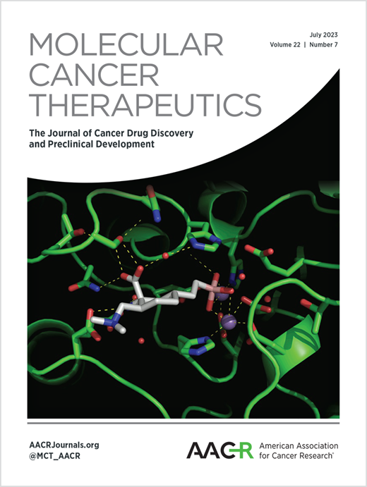
A targeted thorium-227 conjugate demonstrates efficacy in preclinical models of acquired drug resistance and combination potential with chemotherapeutics and antiangiogenic therapies
Molecular Cancer Therapies (2023)
Sabine Zitzmann-Kolbe, Alexander Kristian, Dieter Zopf, Claudia Kamfenkel, Oliver Politz, Christine Ellingsen, Jochen Hilbig, Mark U Juul, Jesper Fonslet, Carsten H Nielsen, Christoph A Schatz, Roger M Bjerke, Alan S Cuthbertson, Dominik Mumberg, Urs B Hagemann
Abstract
Targeted alpha therapies (TATs) are an innovative class of for cancer treatment. The unique mode-of-action of TATs is the induction of deleterious DNA double-strand breaks. Difficult-to-treat cancers, such as gynecological cancers upregulating the chemoresistance P-glycoprotein (p-gp) and overexpressing the membrane protein mesothelin (MSLN), are promising targets for TATs. Here, based on the previous encouraging findings with monotherapy, we investigated the efficacy of the mesothelin-targeted thorium-227 conjugate (MSLN-TTC) both as monotherapy and in combination with chemotherapies and antiangiogenic compounds in ovarian (OvCa) and cervical cancer models expressing p-gp. MSLN-TTC monotherapy showed equal cytotoxicity in vitro in p-gp-positive and negative cancer cells, while chemotherapeutics dramatically lost activity on p-gp-positive cancer cells. In vivo, MSLN-TTC exhibited dose-dependent tumor growth inhibition with T/C ratios of 0.03-0.44 in various xenograft models irrespective of p-gp expression status. Furthermore, MSLN-TTC was more efficacious in p-gp-expressing tumors than chemotherapeutics. In the MSLN-expressing ST206B OvCa PDX model, MSLN-TTC accumulated specifically in the tumor, which combined with pegylated liposomal doxorubicin (doxil), docetaxel, bevacizumab, or regorafenib treatment induced additive-to-synergistic antitumor efficacy and substantially increased response rates compared with respective monotherapies. The combination treatments were well tolerated and only transient decreases in white and red blood cells were observed. In summary, we demonstrate that MSLN-TTC treatment shows efficacy in p-gp-expressing models of chemoresistance and has combination potential with chemo- and antiangiogenic therapies.
Fibroblast activation protein targeted radiotherapy induces an immunogenic tumor microenvironment and enhances the efficacy of PD-1 immune checkpoint inhibition
European Journal of Nuclear Medicine and Molecular Imaging (2023)
Dirk Zboralski, Frank Osterkamp, Esben Christensen, Anne Bredenbeck, Anne Schumann, Aileen Hoehne, Eberhard Schneider, Matthias Paschke, Jan Ungewiss, Christian Haase, Liliane Robillard, Andrew D. Simmons, Thomas C. Harding & Minh Nguyen
Abstract
Purpose: FAP is a membrane-bound protease under investigation as a pan-cancer target, given its high levels in tumors but limited expression in normal tissues. FAP-2286 is a radiopharmaceutical in clinical development for solid tumors that consists of two functional elements: a FAP-targeting peptide and a chelator used to attach radioisotopes. Preclinically, we evaluated the immune modulation and anti-tumor efficacy of FAP-2287, a murine surrogate for FAP-2286, conjugated to the radionuclide lutetium-177 (177Lu) as a monotherapy and in combination with a PD-1 targeting antibody.
Methods: C57BL/6 mice bearing MCA205 mouse FAP-expressing tumors (MCA205-mFAP) were treated with 177Lu-FAP-2287, anti-PD-1, or both. Tumor uptake of 177Lu- FAP-2287 was assessed by SPECT/CT scanning, while therapeutic efficacy was measured by tumor volume and survival. Immune profiling of tumor infiltrates was evaluated through flow cytometry, RNA expression, and immunohistochemistry analyses.
Results: 177Lu-FAP-2287 rapidly accumulated in MCA205-mFAP tumors leading to significant tumor growth inhibition (TGI) and longer survival time. Significant TGI was also observed from anti-PD-1 and the combination. In flow cytometry analysis of tumors, 177Lu-FAP-2287 increased CD8+ T cell infiltration which was maintained in the combination with anti-PD-1. The increase in CD8+ T cells was accompanied by an induction of STING-mediated type I interferon response and higher levels of co-stimulatory molecules such as CD86.
Conclusion: In a preclinical model, FAP-targeted radiotherapy enhanced anti-PD-1-mediated TGI by modulating the TME and increasing the recruitment of tumor-infiltrating CD8+ T cells. These findings provide a rationale for clinical studies of combined 177Lu-FAP-2286 radiotherapy and immune checkpoint inhibition in FAP-positive tumors.

First-in-Human PET Imaging of Tissue Factor in Patients with Primary and Metastatic Cancers Using 18F-labeled Active-Site Inhibited Factor VII (18F-ASIS): Potential as Companion Diagnostic
Journal of Nuclear Medicine, , 2022)
, , , , , , , , , , , , , , and
Abstract
Tissue factor (TF) expression in cancers correlates with poor prognosis. Recently, the first TF-targeted therapy was approved by the U.S. Food and Drug Administration for cervical cancer. To unfold the potential of TF-targeted therapies, correct stratification and selection of patients eligible for treatments may become important for optimization of patient outcomes. TF-targeted PET imaging based on 18F-radiolabeled active-site inhibited versions of the TF natural ligand coagulation factor VII (18F-ASIS) has in preclinical models convincingly demonstrated its use for noninvasive quantitative measurements of TF expression in tumor tissue. 18F-ASIS PET imaging thus has the potential to act as a diagnostic companion for TF-targeted therapies in the clinical setting.
Methods: In this first-in-humans trial, we included 10 cancer patients (4 pancreatic, 3 breast, 2 lung, and 1 cervical cancer) for 18F-ASIS PET imaging. The mean and SD of administered 18F-ASIS activity was 157 ± 35 MBq (range, 93–198 MBq). PET/CT was performed after 1, 2, and 4 h. The primary objectives were to establish the safety, biodistribution, pharmacokinetics, and dosimetry of 18F-ASIS. Secondary objectives included quantitative measurements of SUVs in tumor tissue with PET and evaluation of the correlation (Pearson correlation) between tumor SUVmax and ex vivo TF expression in tumor tissue.
Results: Administration of 18F-ASIS was safe, and no adverse events were observed. No clinically significant changes in vital signs, electrocardiograms, or blood parameters were observed after injection of 18F-ASIS. Mean 18F-ASIS plasma half-life was 3.2 ± 0.6 h, and the radiotracer was predominantly excreted in the urine. For injection activity of 200 MBq of 18F-ASIS, effective whole-body dose was 4 mSv and no prohibitive organ-specific absorbed doses were found. Heterogeneous radiotracer uptake was observed across patients and within tumors. We found a trend of a positive correlation between tumor SUVmax and ex vivo TF expression (r = 0.84, P = 0.08, n = 5).
Conclusion: 18F-ASIS can be safely administered to cancer patients for PET imaging of TF expression in tumors. The trial marks the first test of a TF-targeted PET radiotracer in humans (first-in-class). The findings represent important first steps toward clinical implementation of 18F-ASIS PET imaging of TF expression.
PET in vivo generators 134Ce and 140Nd on an internalizing monoclonal antibody probe
Scientific Reports, 12, (2022)
W. Severin, J. Fonslet, L. K. Kristensen, C. H. Nielsen, A. I. Jensen, A. Kjær, A. P. Mazar, K. Johnston & U. Köster
Abstract
The in vivo-generator radionuclides 140Nd (t1/2 = 3.4 d) and 134Ce (t1/2 = 3.2 d) were used to trace a urokinase-type plasminogen activator (uPA)-targeting mouse monoclonal antibody, ATN-291, in U87 MG xenograft tumor-bearing mice. ATN-291 is known to internalize on the uPA/uPA-receptor pair, making it an appropriate targeting vector for investigating the fate of in vivo generator daughters on internalizing probes. Ante-mortem and post-mortem PET imaging at 120 h post-injection gave no indication of redistribution of the positron emitting daughter nuclides 134La and 140Pr from tumor tissue (p > 0.5). The lack of redistribution indicates that the parent radionuclides 134Ce and 140Nd could be considered as long-lived PET-diagnostic matches to therapeutic radionuclides like 177Lu, 161Tb and 225Ac when internalizing bioconjugates are employed.

The Initial Cardiac Tissue Response to Cryopreserved Allogeneic Adipose Tissue-Derived Mesenchymal Stromal Cells in Rats with Chronic Ischemic Cardiomyopathy
Int. Journal of Molecular Sciences, 22(21), 11758 (2021)
Bjarke Follin, Cecilie Hoeeg, Lisbeth D. Hojgaard, Morten Juhl, Kaya B. Lund, Kristina B. V. Dossing, Simon Bentsen, Ingrid Hunter, Carsten H. Nielsen, Rasmus S. Ripa, Jens Kastrup, Annette Ekblond and Andreas Kjaer
Abstract
Mesenchymal stromal cells have proven capable of improving cardiac pump function in patients with chronic heart failure, yet little is known about their mode of action. The aim of the study was to investigate the short-term effect of cryopreserved allogeneic rat adipose tissue-derived stromal cells (ASC) on cardiac composition, cellular subpopulations, and gene transcription in a rat model of chronic ischemic cardiomyopathy (ICM). Myocardial infarction (MI) was induced by permanent ligation of the left anterior descending coronary artery. After 6 weeks, the rats were treated with ASCs, saline, or no injection, using echo-guided trans-thoracic intramyocardial injections. The cardiac tissue was subsequently collected for analysis of cellular subpopulations and gene transcription 3 and 7 days after treatment. At day 3, an upregulation of genes associated with angiogenesis were present in the ASC group. On day 7, increases in CCR2+ and CD38+ macrophages (p = 0.047 and p = 0.021), as well as in the CD4/CD8 lymphocyte ratio (p = 0.021), were found in the ASC group compared to the saline group. This was supported by an upregulation of genes associated with monocytes/macrophages. In conclusion, ASC treatment initiated an immune response involving monocytes/macrophages and T-cells and induced a gene expression pattern associated with angiogenesis and monocyte/macrophage differentiation.
Immunostimulatory effects of targeted thorium-227 conjugates as single agent and in combination with anti-PD- L1 therapy
Journal for Immunotherapy of Cancer 9, 10 (2021)
Pascale Lejeune, Véronique Cruciani, Axel Berg-Larsen, Andreas Schlicker, Anne Mobergslien, Lisa Bartnitzky, Sandra Berndt, Sabine Zitzmann-Kolbe, Claudia Kamfenkel, Stefan Stargard, Stefanie Hammer, Jennifer S Jørgensen, Malene Jackerott, Carsten H Nielsen, Christoph A Schatz, Hartwig Hennekes, Jenny Karlsson, Alan S Cuthbertson, Dominik Mumberg and Urs B Hagemann
Abstract
Background: Targeted thorium-227 conjugates (TTCs) are an emerging class of targeted alpha therapies (TATs). Their unique mode of action (MoA) is the induction of difficult-to-repair clustered DNA double-strand breaks. However, thus far, their effects on the immune system are largely unknown. Here, we investigated the immunostimulatory effects of the mesothelin-targeted thorium-227 conjugate (MSLN-TTC) in vitro and in vivo in monotherapy and in combination with an inhibitor of the immune checkpoint programmed death receptor ligand 1 (PD-L1) in immunocompetent mice.
Methods: The murine cell line MC38 was transfected with the human gene encoding for MSLN (hMSLN) to enable binding of the non-cross-reactive MSLN-TTC. The immunostimulatory effects of MSLN-TTC were studied in vitro on human cancer cell lines and MC38-hMSLN cells. The efficacy and MoA of MSLN-TTC were studied in vivo as monotherapy or in combination with anti-PD-L1 in MC38-hMSLN tumor-bearing immunocompetent C57BL/6 mice. Experiments were supported by RNA sequencing, flow cytometry, immunohistochemistry, mesoscale, and TaqMan PCR analyses to study the underlying immunostimulatory effects. In vivo depletion of CD8+ T cells and studies with Rag2/Il2Rg double knockout C57BL/6 mice were conducted to investigate the importance of immune cells to the efficacy of MSLN-TTC.
Results: MSLN-TTC treatment induced upregulation of DNA sensing pathway transcripts (IL-6, CCL20, CXCL10, and stimulator of interferon genes (STING)-related genes) in vitro as determined by RNASeq analysis. The results, including phospho-STING activation, were confirmed on the protein level. Danger-associated molecular pattern molecules were upregulated in parallel, leading to dendritic cell (DC) activation in vitro. MSLN-TTC showed strong antitumor activity (T:C 0.38, p<0.05) as a single agent in human MSLN-expressing MC38 tumor-bearing immunocompetent mice. Combining MSLN-TTC with anti-PD-L1 further enhanced the efficacy (T:C 0.08, p<0.001) as evidenced by the increased number of tumor-free surviving animals. MSLN-TTC monotherapy caused migration of CD103+ cDC1 DCs and infiltration of CD8+ T cells into tumors, which was enhanced on combination with anti-PD-L1. Intriguingly, CD8+ T-cell depletion decreased antitumor efficacy.
Conclusions: These in vitro and in vivo data on MSLN-TTC demonstrate that the MoA of TTCs involves activation of the immune system. The findings are of relevance for other targeted radiotherapies and may guide clinical combination strategies.

Darolutamide Potentiates the Antitumor Efficacy of a PSMA-targeted Thorium-227 Conjugate by a Dual Mode of Action in Prostate Cancer Models
Clinical Cancer Research 27 (15): 4367–4378 (2021)
Stefanie Hammer, Andreas Schlicker, Sabine Zitzmann-Kolbe, Simon Baumgart, Urs B. Hagemann, Arne Scholz, Bernard Haendler, Pascale Lejeune, Jenny Karlsson, Christine Ellingsen, Hartwig Hennekes, Carsten H. Nielsen, Mark U. Juul, Dominik Mumberg, and Christoph A. Schatz
Abstract

Preclinical Efficacy of a PSMA-Targeted Thorium-227 Conjugate (PSMA-TTC), a Targeted Alpha Therapy for Prostate Cancer
Clinical Cancer Research 26, 8, 1985–1996 (2020)
Stefanie Hammer, Urs B. Hagemann, Sabine Zitzmann-Kolbe, Aasmund Larsen, Christine Ellingsen, Solene Geraudie, Derek Grant, Baard Indrevoll, Roger Smeets, Oliver von Ahsen, Alexander Kristian, Pascale Lejeune, Hartwig Hennekes, Jenny Karlsson, Roger M. Bjerke, Olav B. Ryan, Alan S. Cuthbertson, and Dominik Mumberg
Abstract:

Monitoring CD8a+ T Cell Responses to Radiotherapy and CTLA-4 Blockade Using [64Cu]NOTA-CD8a PET Imaging
Mol Imaging Biol 22, 1021–1030 (2020)
Kristensen, L.K., Christensen, C., Alfsen, M.Z., Cold, S., Nielsen, C.H., Kjaer, A.
Abstract
Purpose: Current response assessment systems for cancer patients receiving immunotherapy are limited. This is due to the associated inflammatory response that may confound the conventional morphological response evaluation criteria in solid tumors and metabolic positron emission tomography (PET) response criteria in solid. Recently, novel PET imaging techniques using radiolabeled antibodies and fragments have emerged as a particularly sensitive and specific modality for quantitative tracking of immune cell dynamics. Therefore, we sought to investigate the utility of Cu-64 labeled F(ab)′2 fragments for in vivo detection of CD8a+ T cells as a prognostic imaging biomarker of response to immunotherapy in an immunocompetent mouse model of colorectal cancer.
Procedures: [64Cu]NOTA-CD8a was produced by enzymatic digestion of rat-anti-mouse CD8a antibody (clone YTS169.4), purified yielding isolated CD8a-F(ab)′2 fragments and randomly conjugated with the 2-S-(isothiocyanatbenzyl)-1,4,7-triazacyclononane-1,4,7-triacetic acid (p-SCN-Bn-NOTA) chelator. NOTA-CD8a was radiolabeled with Cu-64 and injected into CT26 tumor-bearing mice for longitudinal assessment. To investigate the value of [64Cu]NOTA-CD8a PET imaging for assessment of treatment response, CT26 tumor-bearing mice were subjected to external radiation therapy (XRT) in combination with anti-CTLA-4 therapy. Imaging data was supported by flow cytometry and immunohistochemistry (IHC).
Results: Combination treatment with XRT and anti-CTLA-4 effectively inhibited tumor growth until day 22 post-therapy initiation (p = 0.0025) and increased the overall survival of mice compared to control (p = 0.0017). The [64Cu]NOTA-CD8a tumor-to-heart ratio was increased in XRT + anti-CTLA-4-treated mice on day 8 after initiation of therapy (p = 0.0246). Flow cytometry and IHC confirmed the increase in tumor-infiltrating CD8a+ cells in XRT + anti-CTLA-4-treated mice. Furthermore, [64Cu]NOTA-CD8a PET imaging distinguished responders and non-responders prior to treatment-induced changes in tumor volume among mice.
Conclusion: In the present study, we demonstrated that [64Cu]NOTA-CD8a was able to detect treatment-induced changes in CD8a+ infiltration in murine CT26 colon tumors following a common preclinical combination treatment protocol. Overall, [64Cu]NOTA-CD8a exhibited good prognostic and predictive value. We suggest that [64Cu]NOTA-CD8a PET imaging can be used as an early biomarker of response to therapy in preclinical models.
Quantitative PET imaging of PD-L1 expression in xenograft and syngeneic tumour models using a site-specifically labelled PD-L1 antibody
Eur J Nucl Med Mol Imaging 47, 1302–1313 (2020)
Christensen, C., Kristensen, L.K., Alfsen, M.Z., Nielsen, C.H., Kjaer, A.
Abstract
Purpose: Despite remarkable clinical responses and prolonged survival across several cancers, not all patients benefit from PD-1/PD-L1 immune checkpoint blockade. Accordingly, assessment of tumour PD-L1 expression by immunohistochemistry (IHC) is increasingly applied to guide patient selection, therapeutic monitoring, and improve overall response rates. However, tissue-based methods are invasive and prone to sampling error. We therefore developed a PET radiotracer to specifically detect PD-L1 expression in a non-invasive manner, which could be of diagnostic and predictive value.
Methods: Anti-PD-L1 (clone 6E11, Genentech) was site-specifically conjugated with DIBO-DFO and radiolabelled with 89Zr (89Zr-DFO-6E11). 89Zr-DFO-6E11 was optimized in vivo by longitudinal PET imaging and dose escalation with excess unlabelled 6E11 in HCC827 tumour-bearing mice. Specificity of 89Zr-DFO-6E11 was evaluated in NSCLC xenografts and syngeneic tumour models with different levels of PD-L1 expression. In vivo imaging data was supported by ex vivo biodistribution, flow cytometry, and IHC. To evaluate the predictive value of 89Zr-DFO-6E11 PET imaging, CT26 tumour-bearing mice were subjected to external radiation therapy (XRT) in combination with PD-L1 blockade.
Results: 89Zr-DFO-6E11 was successfully labelled with a high radiochemical purity. The HCC827 tumours and lymphoid tissue were identified by 89Zr-DFO-6E11 PET imaging, and co-injection with 6E11 increased the relative tumour uptake and decreased the splenic uptake. 89Zr-DFO-6E11 detected the differences in PD-L1 expression among tumour models as evaluated by ex vivo methods. 89Zr-DFO-6E11 quantified the increase in PD-L1 expression in tumours and spleens of irradiated mice. XRT and anti-PD-L1 therapy effectively inhibited tumour growth in CT26 tumour-bearing mice (p < 0.01), and the maximum 89Zr-DFO-6E11 tumour-to-muscle ratio correlated with response to therapy (p = 0.0252).
Conclusion: PET imaging with 89Zr-DFO-6E11 is an attractive approach for specific, non-invasive, whole-body visualization of PD-L1 expression. PD-L1 expression can be modulated by radiotherapy regimens and 89Zr-DFO-6E11 PET is able to monitor these changes and predict the response to therapy in an immunocompetent tumour model.
CD4+ and CD8a+ PET imaging predicts response to novel PD-1 checkpoint inhibitor: studies of Sym021 in syngeneic mouse cancer models
Theranostics 9(26), 8221-8238 (2019)
Lotte K. Kristensen, Camilla Fröhlich, Camilla Christensen, Maria C. Melander, Thomas T. Poulsen, Gunther R. Galler, Johan Lantto, Ivan D. Horak, Michael Kragh, Carsten H. Nielsen, Andreas Kjaer
Abstract
Predicting the outcome of immunotherapy is essential for efficient treatment. The recent clinical success of immunotherapy is increasingly changing the paradigm of cancer treatment. Accordingly, the development of immune-based agents is accelerating and the number of agents in the global immuno-oncology pipeline has grown 60-70% over the past year. However, despite remarkable clinical efficacy in some patients, only few achieve a lasting clinical response. Treatment failure can be attributed to poorly immunogenic tumors that do not attract tumor infiltrating lymphocytes (TILs). Therefore, we developed positron emission tomography (PET) radiotracers for non-invasive detection of CD4+ and CD8a+ TILs in syngeneic mouse tumor models for preclinical studies.
Methods: Seven syngeneic mouse tumor models (B16F10, P815, CT26, MC38, Renca, 4T1, Sa1N) were quantified for CD4+ and CD8a+ TILs using flow cytometry and immunohistochemistry (IHC), as well as for tumor growth response to Sym021, a humanized PD-1 antibody cross-reactive with mouse PD-1. Radiotracers were generated from F(ab)’2 fragments of rat-anti-mouse CD4 and CD8a antibodies conjugated to the p-SCN-Bn-Desferrioxamine (SCN-Bn-DFO) chelator and radiolabeled with Zirconium-89 (89Zr-DFO-CD4/89Zr-DFO-CD8a). Tracers were optimized for in vivo PET/CT imaging in CT26 tumor-bearing mice and specificity was evaluated by depletion studies and isotype control imaging. 89Zr-DFO-CD4 and 89Zr-DFO-CD8a PET/CT imaging was conducted in the panel of syngeneic mouse models prior to immunotherapy with Sym021.
Results: Syngeneic tumor models were characterized as “hot” or “cold” according to number of TILs determined by flow cytometry and IHC. 89Zr-DFO-CD4 and 89Zr-DFO-CD8a were successfully generated with a radiochemical purity >99% and immunoreactivity >85%. The optimal imaging time-point was 24 hours post-injection of ~1 MBq tracer with 30 µg non-labeled co-dose. Reduced tumor and spleen uptake of 89Zr-DFO-CD8a was observed in CD8a+ depleted mice and the uptake was comparable with that of isotype control (89Zr-DFO-IgG2b) confirming specificity. PET imaging in syngeneic tumor models revealed a varying maximum tumor-to-heart ratio of 89Zr-DFO-CD4 and 89Zr-DFO-CD8a across tumor types and in-between subjects that correlated with individual response to Sym021 at day 10 relative to start of therapy (p=0.0002 and p=0.0354, respectively). The maximum 89Zr-DFO-CD4 tumor-to-heart ratio could be used to stratify mice according to Sym021 therapy response and overall survival was improved in mice with a 89Zr-DFO-CD4 ratio >9 (p=0.0018). Conclusion: We developed 89Zr-DFO-CD4 and 89Zr-DFO-CD8a PET radiotracers for specific detection and whole-body assessment of CD4+ and CD8a+ status. These radiotracers can be used to phenotype preclinical syngeneic mouse tumor models and to predict response to an immune checkpoint inhibitor. We foresee development of such non-invasive in vivo biomarkers for prediction and evaluation of clinical efficacy of immunotherapeutic agents, such as Sym021.
Mesothelin-Targeted Thorium-227 Conjugate (MSLN-TTC): Preclinical Evaluation of a New Targeted Alpha Therapy for Mesothelin-Positive Cancers
Clininical Cancer Research 25 (15), 4723–4734 (2019)
Urs B. Hagemann, Christine Ellingsen, Joachim Schuhmacher, Alexander Kristian, Anne Mobergslien, Veronique Cruciani, Katrine Wickstroem, Christoph A. Schatz, Christoph Kneip, Sven Golfier, Roger Smeets, Steinar Uran, Hartwig Hennekes, Jenny Karlsson, Roger M. Bjerke, Olav B. Ryan, Dominik Mumberg, Karl Ziegelbauer and Alan S. Cuthbertson
Abstract:
The brain-penetrant clinical ATM inhibitor AZD1390 radiosensitizes and improves survival of preclinical brain tumor models
Science advances 4, 6 (2018)
Stephen T. Durant, Li Zheng, Yingchun Wang, Kan Chen, Lingli Zhang3, Tianwei Zhang, Zhenfan Yang, Lucy Riches, Antonio G. Trinidad, Jacqueline H. L. Fok, Tom Hunt, Kurt G. Pike, Joanne Wilson, Aaron Smith, Nicola Colclough, Venkatesh Pilla Reddy, Andrew Sykes, Annika Janefeldt, Peter Johnstrom, Katarina Varnas, Akihiro Takano, Stephanie Ling, Jonathan Orme, Jonathan Stott, Caroline Roberts, Ian Barrett, Gemma Jones, Martine Roudier, Andrew Pierce, Jasmine Allen, Jenna Kahn, Amrita Sule, Jeremy Karlin, Anna Cronin, Melissa Chapman, Kristoffer Valerie, Ruth Illingworth, Martin Pass
Abstract:
Poor survival rates of patients with tumors arising from or disseminating into the brain are attributed to an inability to excise all tumor tissue (if operable), a lack of blood-brain barrier (BBB) penetration of chemotherapies/targeted agents, and an intrinsic tumor radio-/chemo-resistance. Ataxia-telangiectasia mutated (ATM) protein orchestrates the cellular DNA damage response (DDR) to cytotoxic DNA double-strand breaks induced by ionizing radiation (IR). ATM genetic ablation or pharmacological inhibition results in tumor cell hypersensitivity to IR. We report the primary pharmacology of the clinical-grade, exquisitely potent (cell IC50, 0.78 nM), highly selective [>10,000-fold over kinases within the same phosphatidylinositol 3-kinase-related kinase (PIKK) family], orally bioavailable ATM inhibitor AZD1390 specifically optimized for BBB penetration confirmed in cynomolgus monkey brain positron emission tomography (PET) imaging of microdosed 11C-labeled AZD1390 (Kp,uu, 0.33). AZD1390 blocks ATM-dependent DDR pathway activity and combines with radiation to induce G2 cell cycle phase accumulation, micronuclei, and apoptosis. AZD1390 radiosensitizes glioma and lung cancer cell lines, with p53 mutant glioma cells generally being more radiosensitized than wild type. In in vivo syngeneic and patient-derived glioma as well as orthotopic lung-brain metastatic models, AZD1390 dosed in combination with daily fractions of IR (whole-brain or stereotactic radiotherapy) significantly induced tumor regressions and increased animal survival compared to IR treatment alone. We established a pharmacokinetic-pharmacodynamic-efficacy relationship by correlating free brain concentrations, tumor phospho-ATM/phospho-Rad50 inhibition, apoptotic biomarker (cleaved caspase-3) induction, tumor regression, and survival. On the basis of the data presented here, AZD1390 is now in early clinical development for use as a radiosensitizer in central nervous system malignancies.

PET Imaging of Tissue Factor in Pancreatic Cancer Using 64Cu-Labeled Active Site–Inhibited Factor VII
Journal of Nuclear Medicine, 57 (7) 1112-1119 (2016)
Carsten H. Nielsen, Troels E. Jeppesen, Lotte K. Kristensen, Mette M. Jensen, Henrik H. El Ali, Jacob Madsen, Bo Wiinberg, Lars C. Petersen and Andreas Kjaer
Abstract:
Tissue factor (TF) is the main initiator of the extrinsic coagulation cascade. However, TF also plays an important role in cancer. TF expression has been reported in 53%–89% of all pancreatic adenocarcinomas, and the expression level of TF has in clinical studies correlated with advanced stage, increased microvessel density, metastasis, and poor overall survival. Imaging of TF expression is of clinical relevance as a prognostic biomarker and as a companion diagnostic for TF-directed therapies currently under clinical development. Factor VII (FVII) is the natural ligand to TF. The purpose of this study was to investigate the possibility of using active site–inhibited FVII (FVIIai) labeled with 64Cu for PET imaging of TF expression.
Methods: FVIIai was conjugated to 2-S-(4-isothiocyanatobenzyl)-1,4,7-triazacyclononane-1,4,7-triacetic acid (p-SCN-Bn-NOTA) and labeled with 64Cu (64Cu-NOTA-FVIIai). Longitudinal in vivo PET imaging was performed at 1, 4, 15, and 36 h after injection of 64Cu-NOTA-FVIIai in mice with pancreatic adenocarcinomas (BxPC-3). The specificity of TF imaging with 64Cu-NOTA-FVIIai was investigated in subcutaneous pancreatic tumor models with different levels of TF expression and in a competition experiment. In addition, imaging of orthotopic pancreatic tumors was performed using 64Cu-NOTA-FVIIai and PET/MRI. In vivo imaging data were supported by ex vivo biodistribution, flow cytometry, and immunohistochemistry.
Results: Longitudinal PET imaging with 64Cu-NOTA-FVIIai showed a tumor uptake of 2.3 ± 0.2, 3.7 ± 0.3, 3.4 ± 0.3, and 2.4 ± 0.3 percentage injected dose per gram at 1, 4, 15, and 36 h after injection, respectively. An increase in tumor–to–normal-tissue contrast was observed over the imaging time course. Competition with unlabeled FVIIai significantly (P < 0.001) reduced the tumor uptake. The tumor uptake observed in models with different TF expression levels was significantly different from each other (P < 0.001) and was in agreement with the TF level evaluated by TF immunohistochemistry staining. Orthotopic tumors were clearly visible on the PET/MR images, and the uptake of 64Cu-NOTA-FVIIai was colocalized with viable tumor tissue.
Conclusion: 64Cu-NOTA-FVIIai is well suited for PET imaging of tumor TF expression, and imaging is capable of distinguishing the TF expression level of various pancreatic tumor models.
In vivo imaging of therapy response to a novel Pan-HER antibody mixture using FDG and FLT positron emission tomography
Oncotarget. 2015; 6:37486-37499.
Nielsen C. H., Jensen M. M., Kristensen L. K., Dahlman A., Fröhlich C., Jacobsen H. J., Poulsen T. T., Lantto J., Horak I. D., Kragh M., Kjaer A.
Abstract
Purpose: Overexpression of the human epidermal growth factor receptor (HER) family and their ligands plays an important role in many cancers. Targeting multiple members of the HER family simultaneously may increase the therapeutic efficacy. Here, we report the ability to image the therapeutic response obtained by targeting HER family members individually or simultaneously using the novel monoclonal antibody (mAb) mixture Pan-HER.
Experimental design and results: Mice with subcutaneous BxPC-3 pancreatic adenocarcinomas were divided into five groups receiving vehicle or mAb mixtures directed against either EGFR (HER1), HER2, HER3 or all three receptors combined by Pan-HER. Small animal positron emission tomography/computed tomography (PET/CT) with 2’-deoxy-2’-[18F]fluoro-D-glucose (FDG) and 3’-deoxy-3’-[18F]fluorothymidine (FLT) was performed at baseline and at day 1 or 2 after initiation of therapy. Changes in tumor uptake of tracers were quantified and compared to reduction in tumor size. Imaging results were further validated by immunohistochemistry and qPCR. Mean FDG and FLT uptake in the Pan-HER treated group decreased by 19±4.3% and 24±3.1%, respectively. The early change in FDG and FLT uptake correlated with tumor growth at day 23 relative to day 0. Ex vivo molecular analyses of markers associated with the mechanisms of FDG and FLT uptake confirmed the in vivo imaging results.
Conclusions: Taken together, the study supports the use of FDG and FLT as imaging biomarkers of early response to Pan-HER therapy. FDG and FLT PET/CT imaging should be considered as imaging biomarkers in clinical evaluation of the Pan-HER mAb mixture.
Evaluating the Translation Value of Two In Vivo Models for Breast Cancer Brain Metastases
Cancers (2026)
Abstract
In Vivo Studies of [161Tb]Tb-Trastuzumab Radiopharmaceutical Therapy in Human Epidermal Growth Factor Receptor 2–Expressing Breast Tumors Show High Tumor Uptake and Tumor Growth Suppression
, , , , , , , and
Abstract
Human epidermal growth factor receptor 2 (HER2) has emerged as attractive for targeted radionuclide therapy of HER2-expressing breast cancer. The present study aimed to investigate the image-derived biodistribution, tolerability, and efficacy of [161Tb]Tb-trastuzumab as a potential therapeutic alternative or supplement to trastuzumab and trastuzumab deruxtecan in HER2-expressing tumors. Methods: Trastuzumab was functionalized with DOTA by non–site-specific conjugation and radiolabeled with 161Tb. In vitro assays were performed to evaluate binding affinity and immunoreactivity of [161Tb]Tb-trastuzumab. The influence of mass dose on the biodistribution of [161Tb]Tb-trastuzumab was assessed in mice bearing subcutaneous BT474 tumors by SPECT/CT imaging performed 4, 24, 72, and 168 h after injection of 20 MBq of [161Tb]Tb-trastuzumab labeled at 3 specific activities. Tolerability was evaluated with escalating doses of 5, 10, and 20 MBq of [161Tb]Tb-trastuzumab, and the efficacy of [161Tb]Tb-trastuzumab was compared with that of the vehicle, trastuzumab, and trastuzumab deruxtecan groups. Results: Functionalization and radiolabeling resulted in a radiochemical yield of 97% ± 2% and a radiochemical purity above 99%. The binding affinity of functionalized and native trastuzumab was 0.83 and 0.39 nM, respectively, with an immunoreactive fraction of 89%. SPECT/CT imaging demonstrated peak tumor uptake 72 h after injection, with tumor uptake of 10.8 ± 1.1, 12.8 ± 2.2, and 6.3 ± 0.03 %ID/g of tissue for mice dosed with 0.5, 0.05, and 0.005 MBq/µg, respectively. Radiotoxicity was evident in mice dosed with 20 MBq of [161Tb]Tb-trastuzumab, whereas 5 and 10 MBq of [161Tb]Tb-trastuzumab were well tolerated. Antitumor efficacy was observed in mice dosed with 10 MBq of [161Tb]Tb-trastuzumab, which presented tumor volumes statistically significantly smaller than those of the vehicle group 104 d after treatment (P = 0.0007). Conclusion: Functionalization and radiolabeling did not affect binding affinity. Biodistribution of [161Tb]Tb-trastuzumab revealed high tumor uptake of more than 10 %ID/g of tissue, peaking at 72 h. [161Tb]Tb-trastuzumab in doses of up to 10 MBq was tolerated and highly effective at inhibiting tumor growth (P = 0.0007). These results support the potential of [161Tb]Tb-trastuzumab as an attractive treatment option for HER2-expressing cancers.
A novel synergistic drug combination of a mitogen-activated extracellular signal-regulated kinase inhibitor with [177Lu]Lu-rhPSMA-10.1 for prostate cancer treatment: Results of a preclinical evaluation
Nuclear Medicine and Biology (2025)
, Bart Cornelissen, , Freja Pretzmann, , Mathias Wikke Hallund, and
Abstract
The prostate-specific membrane antigen (PSMA)-targeted radiohybrid ligand [177Lu]Lu-rhPSMA-10.1 is a promising next-generation radiopharmaceutical therapy in prostate cancer. This preclinical evaluation comprised an in vitro screen of potential novel synergistic drug combinations with [177Lu]Lu-rhPSMA-10.1, and an in vivo efficacy analysis of the lead drug combination in PSMA-expressing prostate cancer xenografts. Methods: In total, 177 anticancer drugs were screened in a clonogenic survival assay of 22Rv1 cells which used 5-fold serial dilutions of the test drug (≤ 20 μM) to determine the half-maximal inhibitory concentration (IC50), compared to incubations of the test drug plus [177Lu]Lu-rhPSMA-10.1 (15 MBq) after 10 days. A subsequent focused screen assessed the impact of [177Lu]Lu-rhPSMA-10.1 (0–25 MBq/mL) on drug IC50. Synergy scores were determined using the zero interaction potency (ZIP) reference model (ZIP scores >5 % indicate high synergistic potency) and the multidimensional synergy of combinations (MuSyC) platform (log α >0 indicates synergistic potency). Therapeutic efficacy of the lead drug combination was evaluated in vivo: intravenous [177Lu]Lu-rhPSMA-10.1 (30 MBq, single dose) and oral cobimetinib (0.25 mg/day for 21 days) (alone/in combination) were administered to 22Rv1 tumor-bearing NMRI nude mice (eight mice/group plus untreated controls). Tumor volume was measured twice weekly for 69 days (two-way ANOVA and Tukey’s multiple comparisons test: data analyzed until three mice/group remained). KaplanMeier Log-rank survival analyses were performed. Results: In vitro screening identified cobimetinib (a mitogen-activated extracellular signal-regulated kinase inhibitor) as a lead candidate for synergistic combination with [177Lu]Lu-rhPSMA-10.1 across a wide concentration range (ZIP score=13 %). MuSyC analysis suggested synergistic efficacy from enhanced potency of both drugs in the combination (both log α>3). Combination treatment significantly suppressed tumor growth in vivo versus untreated controls (from Day 13–30; p<0.01) and [177Lu]Lu-rhPSMA-10.1 (from Day 17–30; p<0.001). Median survival was significantly longer with combination treatment (49 days) versus untreated controls (23 days; p=0.001) and [177Lu]Lu-rhPSMA-10.1 monotherapy (36 days; p=0.002). No major compound-related toxicity for cobimetinib ± [177Lu]Lu-rhPSMA-10.1 was observed. Conclusions: The combination of cobimetinib and [177Lu]Lu-rhPSMA-10.1 demonstrated enhanced preclinical therapeutic efficacy versus single agents, supporting clinical investigation of this novel drug combination in prostate cancer.
Preclinical Evaluation of 177Lu-rhPSMA-10.1, a Radiopharmaceutical for Prostate Cancer: Biodistribution and Therapeutic Efficacy
, , , , , and
Abstract
177Lu-rhPSMA-10.1 is a novel radiohybrid prostate-specific membrane antigen (PSMA)–targeted radiopharmaceutical therapy for prostate cancer. We conducted preclinical analyses on non–tumor-bearing BALB/c mice and on prostate cancer human xenograft mouse models (LNCAP and 22Rv1 xenografts) to evaluate its biodistribution and therapeutic efficacy. Methods: Longitudinal biodistribution of 177Lu-rhPSMA-10.1 was evaluated in BALB/c mice and 22Rv1 xenografts. Tissues of interest were harvested, and radioactivity was measured 1–168 h after injection of 1 MBq of 177Lu-rhPSMA-10.1 (4 per time point). Longitudinal biodistribution was compared with 177Lu-PSMA-I&T (1 MBq) in BALB/c mice and at a single time point (15 h) in 22Rv1 xenografts. The therapeutic efficacy of a single administration of 15, 30, or 45 MBq of 177Lu-rhPSMA-10.1 in LNCaP xenografts and 30 MBq of 177Lu-rhPSMA-10.1, 177Lu-PSMA-617, or 177Lu-PSMA-I&T in 22Rv1 xenografts (8 per group) was evaluated. Efficacy versus vehicle was evaluated on the basis of relative tumor volume and survival up to 49 d after administration. Statistical significance was evaluated with t testing (biodistribution data), 2-way repeated-measures ANOVA (tumor volume [analyzed until 3 per group remained]), or Kaplan–Meier log-rank analyses (survival). Results: Biodistribution of 177Lu-rhPSMA-10.1 in the BALB/c and 22Rv1 xenografts showed rapid clearance from blood and other normal tissues within 48 h, with the kidney showing the highest normal-organ uptake. Kidney uptake and retention were lower for 177Lu-rhPSMA-10.1 than for 177Lu-PSMA-I&T (6.5-fold lower at 12 h in BALB/c mice and 6.4-fold lower at 15 h in 22Rv1 xenografts; P < 0.01). High and sustained 177Lu-rhPSMA-10.1 tumor uptake was observed in 22Rv1 xenografts. This uptake was 2.3-fold higher than that of 177Lu-PSMA-I&T (15 h; P < 0.05). When efficacy was evaluated, 177Lu-rhPSMA-10.1 significantly suppressed tumor growth versus vehicle from day 11 (P < 0.05) in LNCaP xenografts in a dose-dependent manner and from day 18 (P < 0.05) in 22Rv1 xenografts and significantly prolonged median survival versus vehicle in both models. In 22Rv1 xenografts, 177Lu-rhPSMA-10.1 suppressed tumor growth versus vehicle to a greater extent than did 177Lu-PSMA-I&T (significant growth inhibition from day 25 [P < 0.05]) and similarly in extent to 177Lu-PSMA-617 (from day 18 [P < 0.05]). Overall, compared with 177Lu-PSMA-I&T, 177Lu-rhPSMA-10.1 suppressed tumor growth for longer than 177Lu-PSMA-617 (inhibition from day 39 onward [P < 0.05] versus on day 49 only [P < 0.05]). Conclusion: In preclinical models, 177Lu-rhPSMA-10.1 shows a favorable tumor-to-kidney uptake ratio, and significant antitumor effects, indicating it to be a promising next-generation radiopharmaceutical therapy.
PET in vivo generators 134Ce and 140Nd on an internalizing monoclonal antibody probe
Scientific Reports, 12, (2022)
W. Severin, J. Fonslet, L. K. Kristensen, C. H. Nielsen, A. I. Jensen, A. Kjær, A. P. Mazar, K. Johnston & U. Köster
Abstract
The in vivo-generator radionuclides 140Nd (t1/2 = 3.4 d) and 134Ce (t1/2 = 3.2 d) were used to trace a urokinase-type plasminogen activator (uPA)-targeting mouse monoclonal antibody, ATN-291, in U87 MG xenograft tumor-bearing mice. ATN-291 is known to internalize on the uPA/uPA-receptor pair, making it an appropriate targeting vector for investigating the fate of in vivo generator daughters on internalizing probes. Ante-mortem and post-mortem PET imaging at 120 h post-injection gave no indication of redistribution of the positron emitting daughter nuclides 134La and 140Pr from tumor tissue (p > 0.5). The lack of redistribution indicates that the parent radionuclides 134Ce and 140Nd could be considered as long-lived PET-diagnostic matches to therapeutic radionuclides like 177Lu, 161Tb and 225Ac when internalizing bioconjugates are employed.
The Initial Cardiac Tissue Response to Cryopreserved Allogeneic Adipose Tissue-Derived Mesenchymal Stromal Cells in Rats with Chronic Ischemic Cardiomyopathy
Int. Journal of Molecular Sciences, 22(21), 11758 (2021)
Bjarke Follin, Cecilie Hoeeg, Lisbeth D. Hojgaard, Morten Juhl, Kaya B. Lund, Kristina B. V. Dossing, Simon Bentsen, Ingrid Hunter, Carsten H. Nielsen, Rasmus S. Ripa, Jens Kastrup, Annette Ekblond and Andreas Kjaer
Abstract
Mesenchymal stromal cells have proven capable of improving cardiac pump function in patients with chronic heart failure, yet little is known about their mode of action. The aim of the study was to investigate the short-term effect of cryopreserved allogeneic rat adipose tissue-derived stromal cells (ASC) on cardiac composition, cellular subpopulations, and gene transcription in a rat model of chronic ischemic cardiomyopathy (ICM). Myocardial infarction (MI) was induced by permanent ligation of the left anterior descending coronary artery. After 6 weeks, the rats were treated with ASCs, saline, or no injection, using echo-guided trans-thoracic intramyocardial injections. The cardiac tissue was subsequently collected for analysis of cellular subpopulations and gene transcription 3 and 7 days after treatment. At day 3, an upregulation of genes associated with angiogenesis were present in the ASC group. On day 7, increases in CCR2+ and CD38+ macrophages (p = 0.047 and p = 0.021), as well as in the CD4/CD8 lymphocyte ratio (p = 0.021), were found in the ASC group compared to the saline group. This was supported by an upregulation of genes associated with monocytes/macrophages. In conclusion, ASC treatment initiated an immune response involving monocytes/macrophages and T-cells and induced a gene expression pattern associated with angiogenesis and monocyte/macrophage differentiation.
Immunostimulatory effects of targeted thorium-227 conjugates as single agent and in combination with anti-PD- L1 therapy
Journal for Immunotherapy of Cancer 9, 10 (2021)
Pascale Lejeune, Véronique Cruciani, Axel Berg-Larsen, Andreas Schlicker, Anne Mobergslien, Lisa Bartnitzky, Sandra Berndt, Sabine Zitzmann-Kolbe, Claudia Kamfenkel, Stefan Stargard, Stefanie Hammer, Jennifer S Jørgensen, Malene Jackerott, Carsten H Nielsen, Christoph A Schatz, Hartwig Hennekes, Jenny Karlsson, Alan S Cuthbertson, Dominik Mumberg and Urs B Hagemann
Abstract
Background: Targeted thorium-227 conjugates (TTCs) are an emerging class of targeted alpha therapies (TATs). Their unique mode of action (MoA) is the induction of difficult-to-repair clustered DNA double-strand breaks. However, thus far, their effects on the immune system are largely unknown. Here, we investigated the immunostimulatory effects of the mesothelin-targeted thorium-227 conjugate (MSLN-TTC) in vitro and in vivo in monotherapy and in combination with an inhibitor of the immune checkpoint programmed death receptor ligand 1 (PD-L1) in immunocompetent mice.
Methods: The murine cell line MC38 was transfected with the human gene encoding for MSLN (hMSLN) to enable binding of the non-cross-reactive MSLN-TTC. The immunostimulatory effects of MSLN-TTC were studied in vitro on human cancer cell lines and MC38-hMSLN cells. The efficacy and MoA of MSLN-TTC were studied in vivo as monotherapy or in combination with anti-PD-L1 in MC38-hMSLN tumor-bearing immunocompetent C57BL/6 mice. Experiments were supported by RNA sequencing, flow cytometry, immunohistochemistry, mesoscale, and TaqMan PCR analyses to study the underlying immunostimulatory effects. In vivo depletion of CD8+ T cells and studies with Rag2/Il2Rg double knockout C57BL/6 mice were conducted to investigate the importance of immune cells to the efficacy of MSLN-TTC.
Results: MSLN-TTC treatment induced upregulation of DNA sensing pathway transcripts (IL-6, CCL20, CXCL10, and stimulator of interferon genes (STING)-related genes) in vitro as determined by RNASeq analysis. The results, including phospho-STING activation, were confirmed on the protein level. Danger-associated molecular pattern molecules were upregulated in parallel, leading to dendritic cell (DC) activation in vitro. MSLN-TTC showed strong antitumor activity (T:C 0.38, p<0.05) as a single agent in human MSLN-expressing MC38 tumor-bearing immunocompetent mice. Combining MSLN-TTC with anti-PD-L1 further enhanced the efficacy (T:C 0.08, p<0.001) as evidenced by the increased number of tumor-free surviving animals. MSLN-TTC monotherapy caused migration of CD103+ cDC1 DCs and infiltration of CD8+ T cells into tumors, which was enhanced on combination with anti-PD-L1. Intriguingly, CD8+ T-cell depletion decreased antitumor efficacy.
Conclusions: These in vitro and in vivo data on MSLN-TTC demonstrate that the MoA of TTCs involves activation of the immune system. The findings are of relevance for other targeted radiotherapies and may guide clinical combination strategies.
Darolutamide Potentiates the Antitumor Efficacy of a PSMA-targeted Thorium-227 Conjugate by a Dual Mode of Action in Prostate Cancer Models
Clinical Cancer Research 27 (15): 4367–4378 (2021)
Stefanie Hammer, Andreas Schlicker, Sabine Zitzmann-Kolbe, Simon Baumgart, Urs B. Hagemann, Arne Scholz, Bernard Haendler, Pascale Lejeune, Jenny Karlsson, Christine Ellingsen, Hartwig Hennekes, Carsten H. Nielsen, Mark U. Juul, Dominik Mumberg, and Christoph A. Schatz
Abstract
Preclinical Efficacy of a PSMA-Targeted Thorium-227 Conjugate (PSMA-TTC), a Targeted Alpha Therapy for Prostate Cancer
Clinical Cancer Research 26, 8, 1985–1996 (2020)
Stefanie Hammer, Urs B. Hagemann, Sabine Zitzmann-Kolbe, Aasmund Larsen, Christine Ellingsen, Solene Geraudie, Derek Grant, Baard Indrevoll, Roger Smeets, Oliver von Ahsen, Alexander Kristian, Pascale Lejeune, Hartwig Hennekes, Jenny Karlsson, Roger M. Bjerke, Olav B. Ryan, Alan S. Cuthbertson, and Dominik Mumberg
Abstract:
Monitoring CD8a+ T Cell Responses to Radiotherapy and CTLA-4 Blockade Using [64Cu]NOTA-CD8a PET Imaging
Mol Imaging Biol 22, 1021–1030 (2020)
Kristensen, L.K., Christensen, C., Alfsen, M.Z., Cold, S., Nielsen, C.H., Kjaer, A.
Abstract
Purpose: Current response assessment systems for cancer patients receiving immunotherapy are limited. This is due to the associated inflammatory response that may confound the conventional morphological response evaluation criteria in solid tumors and metabolic positron emission tomography (PET) response criteria in solid. Recently, novel PET imaging techniques using radiolabeled antibodies and fragments have emerged as a particularly sensitive and specific modality for quantitative tracking of immune cell dynamics. Therefore, we sought to investigate the utility of Cu-64 labeled F(ab)′2 fragments for in vivo detection of CD8a+ T cells as a prognostic imaging biomarker of response to immunotherapy in an immunocompetent mouse model of colorectal cancer.
Procedures: [64Cu]NOTA-CD8a was produced by enzymatic digestion of rat-anti-mouse CD8a antibody (clone YTS169.4), purified yielding isolated CD8a-F(ab)′2 fragments and randomly conjugated with the 2-S-(isothiocyanatbenzyl)-1,4,7-triazacyclononane-1,4,7-triacetic acid (p-SCN-Bn-NOTA) chelator. NOTA-CD8a was radiolabeled with Cu-64 and injected into CT26 tumor-bearing mice for longitudinal assessment. To investigate the value of [64Cu]NOTA-CD8a PET imaging for assessment of treatment response, CT26 tumor-bearing mice were subjected to external radiation therapy (XRT) in combination with anti-CTLA-4 therapy. Imaging data was supported by flow cytometry and immunohistochemistry (IHC).
Results: Combination treatment with XRT and anti-CTLA-4 effectively inhibited tumor growth until day 22 post-therapy initiation (p = 0.0025) and increased the overall survival of mice compared to control (p = 0.0017). The [64Cu]NOTA-CD8a tumor-to-heart ratio was increased in XRT + anti-CTLA-4-treated mice on day 8 after initiation of therapy (p = 0.0246). Flow cytometry and IHC confirmed the increase in tumor-infiltrating CD8a+ cells in XRT + anti-CTLA-4-treated mice. Furthermore, [64Cu]NOTA-CD8a PET imaging distinguished responders and non-responders prior to treatment-induced changes in tumor volume among mice.
Conclusion: In the present study, we demonstrated that [64Cu]NOTA-CD8a was able to detect treatment-induced changes in CD8a+ infiltration in murine CT26 colon tumors following a common preclinical combination treatment protocol. Overall, [64Cu]NOTA-CD8a exhibited good prognostic and predictive value. We suggest that [64Cu]NOTA-CD8a PET imaging can be used as an early biomarker of response to therapy in preclinical models.
Quantitative PET imaging of PD-L1 expression in xenograft and syngeneic tumour models using a site-specifically labelled PD-L1 antibody
Eur J Nucl Med Mol Imaging 47, 1302–1313 (2020)
Christensen, C., Kristensen, L.K., Alfsen, M.Z., Nielsen, C.H., Kjaer, A.
Abstract
Purpose: Despite remarkable clinical responses and prolonged survival across several cancers, not all patients benefit from PD-1/PD-L1 immune checkpoint blockade. Accordingly, assessment of tumour PD-L1 expression by immunohistochemistry (IHC) is increasingly applied to guide patient selection, therapeutic monitoring, and improve overall response rates. However, tissue-based methods are invasive and prone to sampling error. We therefore developed a PET radiotracer to specifically detect PD-L1 expression in a non-invasive manner, which could be of diagnostic and predictive value.
Methods: Anti-PD-L1 (clone 6E11, Genentech) was site-specifically conjugated with DIBO-DFO and radiolabelled with 89Zr (89Zr-DFO-6E11). 89Zr-DFO-6E11 was optimized in vivo by longitudinal PET imaging and dose escalation with excess unlabelled 6E11 in HCC827 tumour-bearing mice. Specificity of 89Zr-DFO-6E11 was evaluated in NSCLC xenografts and syngeneic tumour models with different levels of PD-L1 expression. In vivo imaging data was supported by ex vivo biodistribution, flow cytometry, and IHC. To evaluate the predictive value of 89Zr-DFO-6E11 PET imaging, CT26 tumour-bearing mice were subjected to external radiation therapy (XRT) in combination with PD-L1 blockade.
Results: 89Zr-DFO-6E11 was successfully labelled with a high radiochemical purity. The HCC827 tumours and lymphoid tissue were identified by 89Zr-DFO-6E11 PET imaging, and co-injection with 6E11 increased the relative tumour uptake and decreased the splenic uptake. 89Zr-DFO-6E11 detected the differences in PD-L1 expression among tumour models as evaluated by ex vivo methods. 89Zr-DFO-6E11 quantified the increase in PD-L1 expression in tumours and spleens of irradiated mice. XRT and anti-PD-L1 therapy effectively inhibited tumour growth in CT26 tumour-bearing mice (p < 0.01), and the maximum 89Zr-DFO-6E11 tumour-to-muscle ratio correlated with response to therapy (p = 0.0252).
Conclusion: PET imaging with 89Zr-DFO-6E11 is an attractive approach for specific, non-invasive, whole-body visualization of PD-L1 expression. PD-L1 expression can be modulated by radiotherapy regimens and 89Zr-DFO-6E11 PET is able to monitor these changes and predict the response to therapy in an immunocompetent tumour model.
CD4+ and CD8a+ PET imaging predicts response to novel PD-1 checkpoint inhibitor: studies of Sym021 in syngeneic mouse cancer models
Theranostics 9(26), 8221-8238 (2019)
Lotte K. Kristensen, Camilla Fröhlich, Camilla Christensen, Maria C. Melander, Thomas T. Poulsen, Gunther R. Galler, Johan Lantto, Ivan D. Horak, Michael Kragh, Carsten H. Nielsen, Andreas Kjaer
Abstract
Predicting the outcome of immunotherapy is essential for efficient treatment. The recent clinical success of immunotherapy is increasingly changing the paradigm of cancer treatment. Accordingly, the development of immune-based agents is accelerating and the number of agents in the global immuno-oncology pipeline has grown 60-70% over the past year. However, despite remarkable clinical efficacy in some patients, only few achieve a lasting clinical response. Treatment failure can be attributed to poorly immunogenic tumors that do not attract tumor infiltrating lymphocytes (TILs). Therefore, we developed positron emission tomography (PET) radiotracers for non-invasive detection of CD4+ and CD8a+ TILs in syngeneic mouse tumor models for preclinical studies.
Methods: Seven syngeneic mouse tumor models (B16F10, P815, CT26, MC38, Renca, 4T1, Sa1N) were quantified for CD4+ and CD8a+ TILs using flow cytometry and immunohistochemistry (IHC), as well as for tumor growth response to Sym021, a humanized PD-1 antibody cross-reactive with mouse PD-1. Radiotracers were generated from F(ab)’2 fragments of rat-anti-mouse CD4 and CD8a antibodies conjugated to the p-SCN-Bn-Desferrioxamine (SCN-Bn-DFO) chelator and radiolabeled with Zirconium-89 (89Zr-DFO-CD4/89Zr-DFO-CD8a). Tracers were optimized for in vivo PET/CT imaging in CT26 tumor-bearing mice and specificity was evaluated by depletion studies and isotype control imaging. 89Zr-DFO-CD4 and 89Zr-DFO-CD8a PET/CT imaging was conducted in the panel of syngeneic mouse models prior to immunotherapy with Sym021.
Results: Syngeneic tumor models were characterized as “hot” or “cold” according to number of TILs determined by flow cytometry and IHC. 89Zr-DFO-CD4 and 89Zr-DFO-CD8a were successfully generated with a radiochemical purity >99% and immunoreactivity >85%. The optimal imaging time-point was 24 hours post-injection of ~1 MBq tracer with 30 µg non-labeled co-dose. Reduced tumor and spleen uptake of 89Zr-DFO-CD8a was observed in CD8a+ depleted mice and the uptake was comparable with that of isotype control (89Zr-DFO-IgG2b) confirming specificity. PET imaging in syngeneic tumor models revealed a varying maximum tumor-to-heart ratio of 89Zr-DFO-CD4 and 89Zr-DFO-CD8a across tumor types and in-between subjects that correlated with individual response to Sym021 at day 10 relative to start of therapy (p=0.0002 and p=0.0354, respectively). The maximum 89Zr-DFO-CD4 tumor-to-heart ratio could be used to stratify mice according to Sym021 therapy response and overall survival was improved in mice with a 89Zr-DFO-CD4 ratio >9 (p=0.0018). Conclusion: We developed 89Zr-DFO-CD4 and 89Zr-DFO-CD8a PET radiotracers for specific detection and whole-body assessment of CD4+ and CD8a+ status. These radiotracers can be used to phenotype preclinical syngeneic mouse tumor models and to predict response to an immune checkpoint inhibitor. We foresee development of such non-invasive in vivo biomarkers for prediction and evaluation of clinical efficacy of immunotherapeutic agents, such as Sym021.
Mesothelin-Targeted Thorium-227 Conjugate (MSLN-TTC): Preclinical Evaluation of a New Targeted Alpha Therapy for Mesothelin-Positive Cancers
Clininical Cancer Research 25 (15), 4723–4734 (2019)
Urs B. Hagemann, Christine Ellingsen, Joachim Schuhmacher, Alexander Kristian, Anne Mobergslien, Veronique Cruciani, Katrine Wickstroem, Christoph A. Schatz, Christoph Kneip, Sven Golfier, Roger Smeets, Steinar Uran, Hartwig Hennekes, Jenny Karlsson, Roger M. Bjerke, Olav B. Ryan, Dominik Mumberg, Karl Ziegelbauer and Alan S. Cuthbertson
Abstract:
The brain-penetrant clinical ATM inhibitor AZD1390 radiosensitizes and improves survival of preclinical brain tumor models
Science advances 4, 6 (2018)
Stephen T. Durant, Li Zheng, Yingchun Wang, Kan Chen, Lingli Zhang3, Tianwei Zhang, Zhenfan Yang, Lucy Riches, Antonio G. Trinidad, Jacqueline H. L. Fok, Tom Hunt, Kurt G. Pike, Joanne Wilson, Aaron Smith, Nicola Colclough, Venkatesh Pilla Reddy, Andrew Sykes, Annika Janefeldt, Peter Johnstrom, Katarina Varnas, Akihiro Takano, Stephanie Ling, Jonathan Orme, Jonathan Stott, Caroline Roberts, Ian Barrett, Gemma Jones, Martine Roudier, Andrew Pierce, Jasmine Allen, Jenna Kahn, Amrita Sule, Jeremy Karlin, Anna Cronin, Melissa Chapman, Kristoffer Valerie, Ruth Illingworth, Martin Pass
Abstract:
Poor survival rates of patients with tumors arising from or disseminating into the brain are attributed to an inability to excise all tumor tissue (if operable), a lack of blood-brain barrier (BBB) penetration of chemotherapies/targeted agents, and an intrinsic tumor radio-/chemo-resistance. Ataxia-telangiectasia mutated (ATM) protein orchestrates the cellular DNA damage response (DDR) to cytotoxic DNA double-strand breaks induced by ionizing radiation (IR). ATM genetic ablation or pharmacological inhibition results in tumor cell hypersensitivity to IR. We report the primary pharmacology of the clinical-grade, exquisitely potent (cell IC50, 0.78 nM), highly selective [>10,000-fold over kinases within the same phosphatidylinositol 3-kinase-related kinase (PIKK) family], orally bioavailable ATM inhibitor AZD1390 specifically optimized for BBB penetration confirmed in cynomolgus monkey brain positron emission tomography (PET) imaging of microdosed 11C-labeled AZD1390 (Kp,uu, 0.33). AZD1390 blocks ATM-dependent DDR pathway activity and combines with radiation to induce G2 cell cycle phase accumulation, micronuclei, and apoptosis. AZD1390 radiosensitizes glioma and lung cancer cell lines, with p53 mutant glioma cells generally being more radiosensitized than wild type. In in vivo syngeneic and patient-derived glioma as well as orthotopic lung-brain metastatic models, AZD1390 dosed in combination with daily fractions of IR (whole-brain or stereotactic radiotherapy) significantly induced tumor regressions and increased animal survival compared to IR treatment alone. We established a pharmacokinetic-pharmacodynamic-efficacy relationship by correlating free brain concentrations, tumor phospho-ATM/phospho-Rad50 inhibition, apoptotic biomarker (cleaved caspase-3) induction, tumor regression, and survival. On the basis of the data presented here, AZD1390 is now in early clinical development for use as a radiosensitizer in central nervous system malignancies.
PET Imaging of Tissue Factor in Pancreatic Cancer Using 64Cu-Labeled Active Site–Inhibited Factor VII
Journal of Nuclear Medicine, 57 (7) 1112-1119 (2016)
Carsten H. Nielsen, Troels E. Jeppesen, Lotte K. Kristensen, Mette M. Jensen, Henrik H. El Ali, Jacob Madsen, Bo Wiinberg, Lars C. Petersen and Andreas Kjaer
Abstract:
Tissue factor (TF) is the main initiator of the extrinsic coagulation cascade. However, TF also plays an important role in cancer. TF expression has been reported in 53%–89% of all pancreatic adenocarcinomas, and the expression level of TF has in clinical studies correlated with advanced stage, increased microvessel density, metastasis, and poor overall survival. Imaging of TF expression is of clinical relevance as a prognostic biomarker and as a companion diagnostic for TF-directed therapies currently under clinical development. Factor VII (FVII) is the natural ligand to TF. The purpose of this study was to investigate the possibility of using active site–inhibited FVII (FVIIai) labeled with 64Cu for PET imaging of TF expression.
Methods: FVIIai was conjugated to 2-S-(4-isothiocyanatobenzyl)-1,4,7-triazacyclononane-1,4,7-triacetic acid (p-SCN-Bn-NOTA) and labeled with 64Cu (64Cu-NOTA-FVIIai). Longitudinal in vivo PET imaging was performed at 1, 4, 15, and 36 h after injection of 64Cu-NOTA-FVIIai in mice with pancreatic adenocarcinomas (BxPC-3). The specificity of TF imaging with 64Cu-NOTA-FVIIai was investigated in subcutaneous pancreatic tumor models with different levels of TF expression and in a competition experiment. In addition, imaging of orthotopic pancreatic tumors was performed using 64Cu-NOTA-FVIIai and PET/MRI. In vivo imaging data were supported by ex vivo biodistribution, flow cytometry, and immunohistochemistry.
Results: Longitudinal PET imaging with 64Cu-NOTA-FVIIai showed a tumor uptake of 2.3 ± 0.2, 3.7 ± 0.3, 3.4 ± 0.3, and 2.4 ± 0.3 percentage injected dose per gram at 1, 4, 15, and 36 h after injection, respectively. An increase in tumor–to–normal-tissue contrast was observed over the imaging time course. Competition with unlabeled FVIIai significantly (P < 0.001) reduced the tumor uptake. The tumor uptake observed in models with different TF expression levels was significantly different from each other (P < 0.001) and was in agreement with the TF level evaluated by TF immunohistochemistry staining. Orthotopic tumors were clearly visible on the PET/MR images, and the uptake of 64Cu-NOTA-FVIIai was colocalized with viable tumor tissue.
Conclusion: 64Cu-NOTA-FVIIai is well suited for PET imaging of tumor TF expression, and imaging is capable of distinguishing the TF expression level of various pancreatic tumor models.
In vivo imaging of therapy response to a novel Pan-HER antibody mixture using FDG and FLT positron emission tomography
Oncotarget. 2015; 6:37486-37499.
Nielsen C. H., Jensen M. M., Kristensen L. K., Dahlman A., Fröhlich C., Jacobsen H. J., Poulsen T. T., Lantto J., Horak I. D., Kragh M., Kjaer A.
Abstract
Purpose: Overexpression of the human epidermal growth factor receptor (HER) family and their ligands plays an important role in many cancers. Targeting multiple members of the HER family simultaneously may increase the therapeutic efficacy. Here, we report the ability to image the therapeutic response obtained by targeting HER family members individually or simultaneously using the novel monoclonal antibody (mAb) mixture Pan-HER.
Experimental design and results: Mice with subcutaneous BxPC-3 pancreatic adenocarcinomas were divided into five groups receiving vehicle or mAb mixtures directed against either EGFR (HER1), HER2, HER3 or all three receptors combined by Pan-HER. Small animal positron emission tomography/computed tomography (PET/CT) with 2’-deoxy-2’-[18F]fluoro-D-glucose (FDG) and 3’-deoxy-3’-[18F]fluorothymidine (FLT) was performed at baseline and at day 1 or 2 after initiation of therapy. Changes in tumor uptake of tracers were quantified and compared to reduction in tumor size. Imaging results were further validated by immunohistochemistry and qPCR. Mean FDG and FLT uptake in the Pan-HER treated group decreased by 19±4.3% and 24±3.1%, respectively. The early change in FDG and FLT uptake correlated with tumor growth at day 23 relative to day 0. Ex vivo molecular analyses of markers associated with the mechanisms of FDG and FLT uptake confirmed the in vivo imaging results.
Conclusions: Taken together, the study supports the use of FDG and FLT as imaging biomarkers of early response to Pan-HER therapy. FDG and FLT PET/CT imaging should be considered as imaging biomarkers in clinical evaluation of the Pan-HER mAb mixture.